⚡ The Problem: Every Minute Counts
Every year, millions of MRI scans are performed worldwide, with a large portion involving neurological conditions. Demand is growing faster than the availability of neuroradiology services, causing staffing shortages, diagnostic delays, and errors. Depending on where the scan is performed, results can be delayed by days or even longer.
This is particularly critical for conditions where time literally counts in minutes. An ischemic stroke requires treatment within 4.5 hours — and every minute of delay destroys approximately 1.9 million neurons. A cerebral hemorrhage can become fatal within hours. In these cases, an AI that can immediately prioritize critical cases isn't a luxury — it's a necessity.
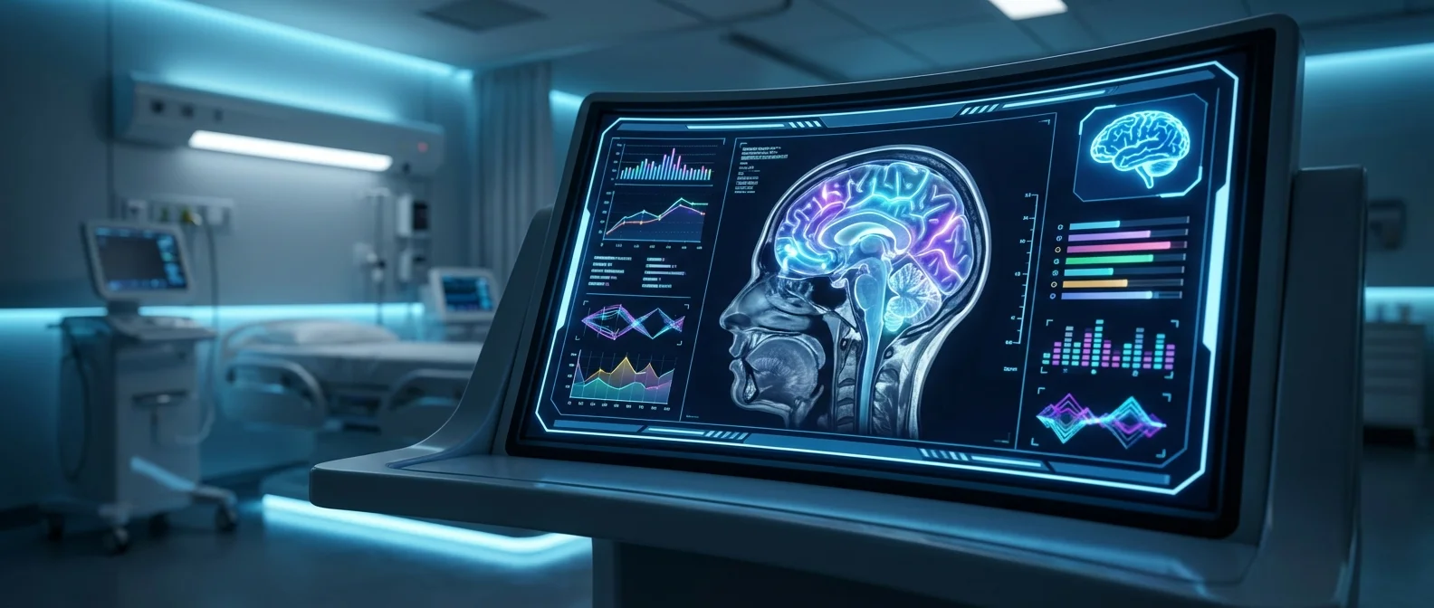
🧠 Prima: “ChatGPT for Medical Imaging”
Researchers at the University of Michigan, led by neurosurgeon Todd Hollon, developed the Prima system — a vision language model (VLM) that processes images, video, and text simultaneously in real time. It's not just another AI that searches for a specific condition in a scan. It's an AI that reads the entire scan like an experienced radiologist — taking into account the patient's medical history and the reason the scan was ordered.
What sets Prima apart from previous AI systems in radiology is the breadth of its data. Most previous models were trained on carefully selected samples and were designed for narrow tasks — detecting lesions or estimating dementia risk. Prima was trained on every available MRI that had been digitized at University of Michigan Health: 200,000+ MRI studies and 5.6 million imaging sequences.
"Just as an AI tool can help draft an email or suggest recommendations, Prima aims to be a co-pilot for interpreting medical imaging studies."
— Todd Hollon, M.D., University of Michigan
📊 97.5% Accuracy Across 50+ Diagnoses
Prima was evaluated on over 30,000 MRI studies over a one-year period. Across more than 50 different radiological diagnoses — tumors, hemorrhages, ischemic strokes, neurodegenerative diseases — the system achieved accuracy up to 97.5%, surpassing other advanced AI models.
Beyond diagnosis, Prima has a capability that makes it particularly valuable in practice: it assesses the level of urgency. It doesn't just identify the problem — it determines whether immediate action is needed and automatically notifies the appropriate specialist: a stroke neurologist for a stroke, a neurosurgeon for a tumor. Feedback becomes available immediately after the scan is completed.
📊 Prima in Numbers
- 200,000+ MRI studies in the training set
- 5.6 million imaging sequences and clinical data
- 97.5% accuracy across 50+ diagnoses
- 30,000+ MRIs evaluated over 12 months
- Seconds instead of hours or days of waiting
🏥 Why This Isn't Just Another AI
Prima differs from most AI systems in healthcare in one critical way: it operates like a real radiologist. A radiologist doesn't just look at the image — they combine the image with the medical history, symptoms, and the reason for the examination. Prima does exactly that: it integrates clinical history and referral reasons along with imaging data to produce comprehensive understanding.
"Prima operates like a radiologist, integrating information about the patient's medical history and imaging data to produce comprehensive understanding of their health," explained Samir Harake, a data scientist in Hollon's lab.
🌍 The Big Picture: Health Equity
Beyond large hospitals, Prima holds particular significance for resource-limited areas. "Whether you're scanning at a large health system facing increasing volume or at a rural hospital with limited resources, innovative technologies are needed to improve access to radiology services," said Vikas Gulani, chair of the U-M Health Department of Radiology.
The researchers emphasize that the system is still in an early evaluation phase. Future research will focus on integrating more detailed patient information and electronic health record data. But Hollon sees further ahead: similar technology could be applied to mammograms, chest X-rays, and ultrasounds — transforming how medical imaging is done worldwide.
The study was published in Nature Biomedical Engineering in February 2026 and was funded by the NIH, the Chan Zuckerberg Initiative, and the Frankel Institute for Heart and Brain Health.