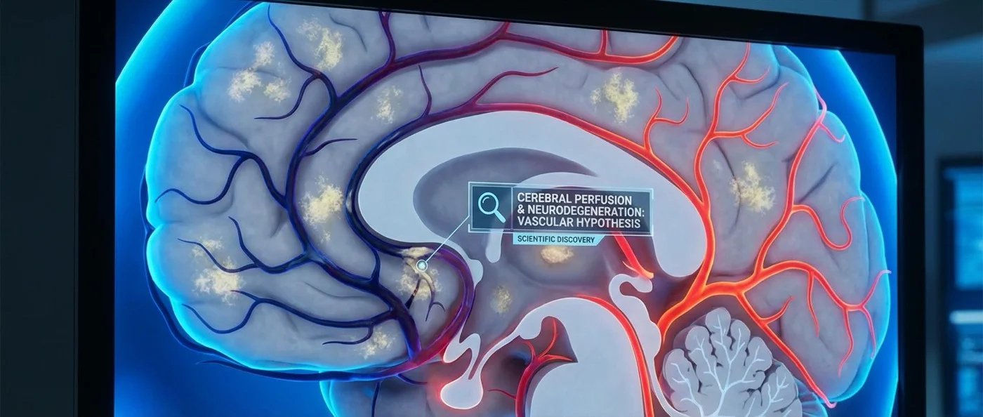
Alzheimer's disease is typically described as a protein problem — amyloid plaques and tau tangles building up in the brain over decades. But a new study from USC's Keck School of Medicine refocuses attention on a less-discussed factor: blood flow. Using two inexpensive, noninvasive tools, researchers showed that measurable declines in brain blood flow and oxygen delivery are closely linked to amyloid accumulation and hippocampal shrinkage — and may be detectable years before any symptoms appear.
📖 Read more: Viagra & Shingles Vaccine Cut Alzheimer's Risk by 17%
🧠 Blood Flow and Oxygen: Alzheimer's Overlooked Risk Indicators
Lead researcher Amaryllis Tsiknia and senior researcher Meredith Braskie from the USC Mark and Mary Stevens Neuroimaging and Informatics Institute set out to test whether simple vascular measurements could identify Alzheimer's risk early — without the cost, radiation, or inaccessibility of PET scans and MRIs.
Their study, published in Alzheimer's & Dementia in February 2026, evaluated participants across three cognitive groups: cognitively normal adults, those with mild cognitive impairment (MCI), and those with full dementia. All underwent vascular function measurements using two complementary tools.
🔬 The Two Tools: TCD and NIRS
The study combined measurements from two established but underutilized technologies:
- Transcranial Doppler Ultrasound (TCD): Measures the speed of blood flow in the brain's major arteries using sound waves — no radiation, no contrast agents. It works similarly to the ultrasound used in prenatal care, but focuses on the cerebral arteries.
- Near-Infrared Spectroscopy (NIRS): Uses harmless near-infrared light that passes through the skull to measure how much oxygen is actively being delivered to the brain cortex in real time.
By combining readings from both devices in mathematical models, the team generated composite indices of cerebrovascular function for each participant. These indices were then cross-referenced with amyloid burden, hippocampal volume, and cognitive status.
🔑 Core Finding
Better cerebrovascular function = lower amyloid burden + larger hippocampus = lower Alzheimer's risk. The hippocampus — the brain's memory center — is among the first regions damaged by Alzheimer's, and its volume loss is one of the earliest measurable signs. Vascular health appears to be directly linked to this pathway.
🏥 What the Study Found: Comparing Patient Groups
As expected, patients with MCI and dementia showed significantly weaker vascular function scores compared to cognitively normal participants. The blood flow and oxygen delivery to their brains was measurably reduced.
The key finding was more nuanced: even among cognitively normal participants, those who had elevated amyloid levels (a preclinical Alzheimer's indicator) showed significantly lower vascular function scores than those without amyloid accumulation. This means the tools may be able to flag risk during the silent, pre-symptomatic phase of the disease — years before memory problems emerge.
💡 Why This Matters: Democratizing Alzheimer's Detection
Current gold-standard Alzheimer's diagnostics — amyloid PET scans and cerebrospinal fluid analysis — are expensive, invasive, and largely inaccessible outside major medical centers. In much of the world, these tools simply aren't available. In the United States, a single amyloid PET scan costs thousands of dollars and requires specialized equipment.
Transcranial Doppler and NIRS devices are inexpensive, portable, and can be used in community clinic settings. If validated in larger multi-site longitudinal studies, this approach could make early Alzheimer's screening accessible to millions of people globally — particularly in low- and middle-income countries where the burden of dementia is growing fastest.
"Amyloid and tau are often considered the primary players in Alzheimer's disease, but blood flow and oxygen delivery to the brain are also critical — and they can be measured simply and non-invasively."
— Amaryllis Tsiknia, PhD, USC Keck School of Medicine, Alzheimer's & Dementia 2026🔭 Limitations and Next Steps
This is a cross-sectional study — it captures a single snapshot in time, not patients followed across years. To establish that vascular decline predicts Alzheimer's, longitudinal studies are needed that follow participants from healthy baseline to eventual cognitive decline. These are now underway at USC.
The study was funded by NIH grants S10OD032285 and R01AG058162, with senior authorship from Arthur Toga, director of the USC Stevens Neuroimaging and Informatics Institute. The findings represent a meaningful step toward simpler, more accessible Alzheimer's risk assessment that doesn't require a PET scanner.