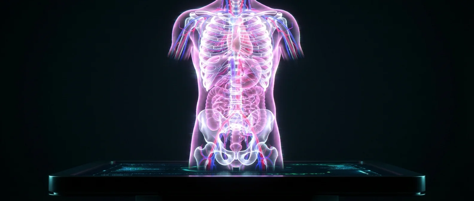
An ultrasound shows you the structure. An MRI maps your tissues. But neither shows you the form and the function of the body simultaneously — in 3D, in color, without radiation, in less than a minute. Until now. Researchers at Caltech and USC developed a technique that does exactly that, just published in Nature Biomedical Engineering.
🔬 Why Today's Tools Are Not Enough
Medical imaging has come a long way from the first black-and-white X-rays. MRIs, CT scans, PET scans — all provide valuable information. But each technology has significant drawbacks.
A conventional ultrasound is fast, cheap, and widely available, but it mainly shows the shape of tissues in two dimensions and covers a limited area. CT scans expose the patient to ionizing radiation. MRI is expensive, time-consuming, and often requires contrast agents. What they all have in common: none of them simultaneously shows tissue structure and blood vessel function in a single, three-dimensional color map.
Photoacoustic imaging offers a different perspective: it sends laser pulses into the tissue, and molecules that absorb light produce sound waves — the so-called photoacoustic signals. These reveal blood vessels in optical color, allowing doctors to distinguish arteries from veins. However, photoacoustic imaging doesn't show tissue structure well.
💡 The Solution: RUS-PAT
Lihong Wang, professor of Medical Engineering and Electrical Engineering at Caltech and a pioneer in photoacoustic tomography for over two decades, asked: why not combine both? Ultrasound for structure, photoacoustic tomography for function — in a single system.
The challenge was technical. Conventional ultrasound systems use multiple transducers to send and receive sound waves, making integration with photoacoustic imaging complex and expensive. Wang found an elegant solution: instead of multiple ultrasound transmitters, a single wide-field transmitter can send sound waves across all the tissue — an approach inspired by the way light diffuses in photoacoustic technique.
The result was named RUS-PAT (Rotational Ultrasound Tomography, RUST, combined with Photoacoustic Tomography, PAT). Arc-shaped detectors rotate around a central point, functioning like a full hemispherical detector — but much simpler and cheaper.
How It Works
Laser pulses are sent into the tissue. Hemoglobin molecules in the blood absorb the light and emit sound waves. Simultaneously, a wide-field ultrasound transmitter sends sound waves into the tissue. The same detectors capture both signals, creating an image that shows structure in 3D (from the ultrasound) and function in color (from the photoacoustic).
🏥 What It Can Do in Practice
The technology isn't theoretical — it has already been tested on humans. Across various parts of the body, RUS-PAT produced images that simultaneously show soft tissues (muscles, fat, skin) and blood vessels in full color resolution.
The researchers identify three main areas of clinical application. In breast imaging, RUS-PAT can detect tumors showing not only their location but also their biological activity — without radiation or contrast agents. For patients with diabetic neuropathy, the technique allows simultaneous monitoring of both nerve structure and oxygen supply in a single scan. Finally, in brain research, scientists can study anatomy while simultaneously observing blood flow.
🧪 The Big Picture: From CERN to Caltech
RUS-PAT isn't the only effort to bring color to medical imaging. In 2018, Mars Bioimaging from New Zealand unveiled the first three-dimensional color X-ray scanner, using a Medipix3 chip originally developed at CERN for the Large Hadron Collider. The technology, called Spectral CT, measures the attenuation of specific wavelengths of X-rays as they pass through different materials, producing images that clearly separate bone, muscle, fat, vessels, and disease markers.
That same year, researchers at UC Davis unveiled EXPLORER — the world's first full-body PET/CT scanner. Nearly 40 times more sensitive than existing systems, EXPLORER can scan the entire body in 20-30 seconds instead of 40 minutes, with significantly lower radiation doses.
The trend is clear: medical imaging is moving toward faster, safer, more detailed scans. Caltech's RUS-PAT stands out because it achieves something no other system offers: structure and function simultaneously, without radiation, in less than a minute.
⚕️ What It Means for Patients
"The new combination of acoustic and photoacoustic techniques addresses many of the key limitations of widely used medical imaging techniques," said Dr. Charles Y. Liu, co-author of the study, professor at USC's Keck School of Medicine, and director of the Neurorestoration Center. "And, crucially, its applicability to humans has already been demonstrated in multiple settings."
For patients, the advantages are obvious. No radiation, no contrast agents, no need for expensive MRI equipment. A scan under one minute instead of half-hour or hour-long procedures. And images that don't just show “what tissue is there,” but “how it's functioning right now” — whether vessels are carrying enough oxygen, whether a tumor is actively being fed, whether nerves are receiving proper blood supply.
The current setup places the ultrasound transmitters and laser beneath a scanning bed. The system has already been tested on volunteers and patients and is in the early stages of transitioning to clinical use. Additionally, light can be delivered through endoscopic tools, which could enable access to deeper areas of the body in the future.
"It's not about one plus one," explains Wang. “We needed to find the optimal way to combine two technologies.” That optimal approach can now see the human body in three dimensions, in color, in real time — and medicine will never be the same.